
")
Advancing Precision Medicine: NCKU Researchers’ Innovation in Disc Herniation Detection on Cover Story of Journal of Biomedical Materials Research Part A
Written by Hsu Tsu-Yueh. Image credit to NCKU News Center and Yeau-Ren Jeng

Chair Professor Yeau-Ren Jeng from NCKU Department of Biomedical Engineering (middle). Team member Azril, Ph.D. student of NCKU Department of Biomedical Engineering (right). Attending Physician Kuo-Yuan Huang from NCKU Hospital Department of Orthopedics (left).
Chair Professor Yeau-Ren Jeng (鄭友仁) from the Department of Biomedical Engineering at National Cheng Kung University and Attending Physician Kuo-Yuan Huang (黃國淵) from the Department of Orthopedics at NCKU hospital collaborated to innovate a system integrating nanoindentation with Raman spectrometry to achieve in-depth analysis of the internal condition of intervertebral discs.
Through precision testing, they gained insights into the state of the nucleus pulposus in the intervertebral discs, allowing for an analysis of the degree of degeneration and inflammation. This enables doctors to understand the severity of the condition and determine whether conservative or surgical treatment is appropriate for the patients.
The research was published in the official journal of the International Society for Biomaterials, the Journal of Biomedical Materials Research Part A, and was selected as the cover story in July. (Article link: Correlation of the degenerative stage of a disc with magnetic resonance imaging, chemical content, and biomechanical properties of the nucleus pulposus)
The research was published in the official journal of the International Society for Biomaterials, the Journal of Biomedical Materials Research and was selected as the cover story.
Dr. Kuo-Yuan Huang stated that spinal degeneration is a common condition that affects many people worldwide. When the spine becomes unstable, one may develop spondylolisthesis or scoliosis, leading to back pain, sciatica, and limping symptoms. Additionally, it may compress vital nerve structures, with disc herniation being one of the common diseases.
The intervertebral disc is a cartilaginous tissue, with the outer ring called annulus fibrosus and the inner core called nucleus pulposus (NP), which resembles a jelly-like texture. When the outer annulus fibrosus wears down and cracks appear, the “jelly” can be squeezed out from the crack, compressing the nearby nervous system.
Patients may experience numbness, tingling, or pain in the lower back or limbs, which is sciatica caused by disc herniation. Disc herniation is more common among laborers or sedentary office workers. Suppose one lifts heavy objects or bends over for a long time at work, with increasing usage and age. In that case, the working conditions can also lead to the loss of elasticity in the “jelly,” causing NP herniation, which compresses nerves and may eventually lead to intervertebral disc degeneration (IDD).
“Magnetic Resonance Imaging (MRI) is currently the primary tool doctors use to detect the degree of spinal degeneration. Yet, MRI can only provide diagnostic judgments based on images," said Dr. Kuo-Yuan Huang. Currently, the method for detecting disc herniation relies on MRI. However, through image interpretation, it is impossible to discern the internal components, understand specific changes in the properties such as the weight-bearing ability of the disc and the chemical composition of the nucleus pulposus, or obtain more detailed mechanical information.
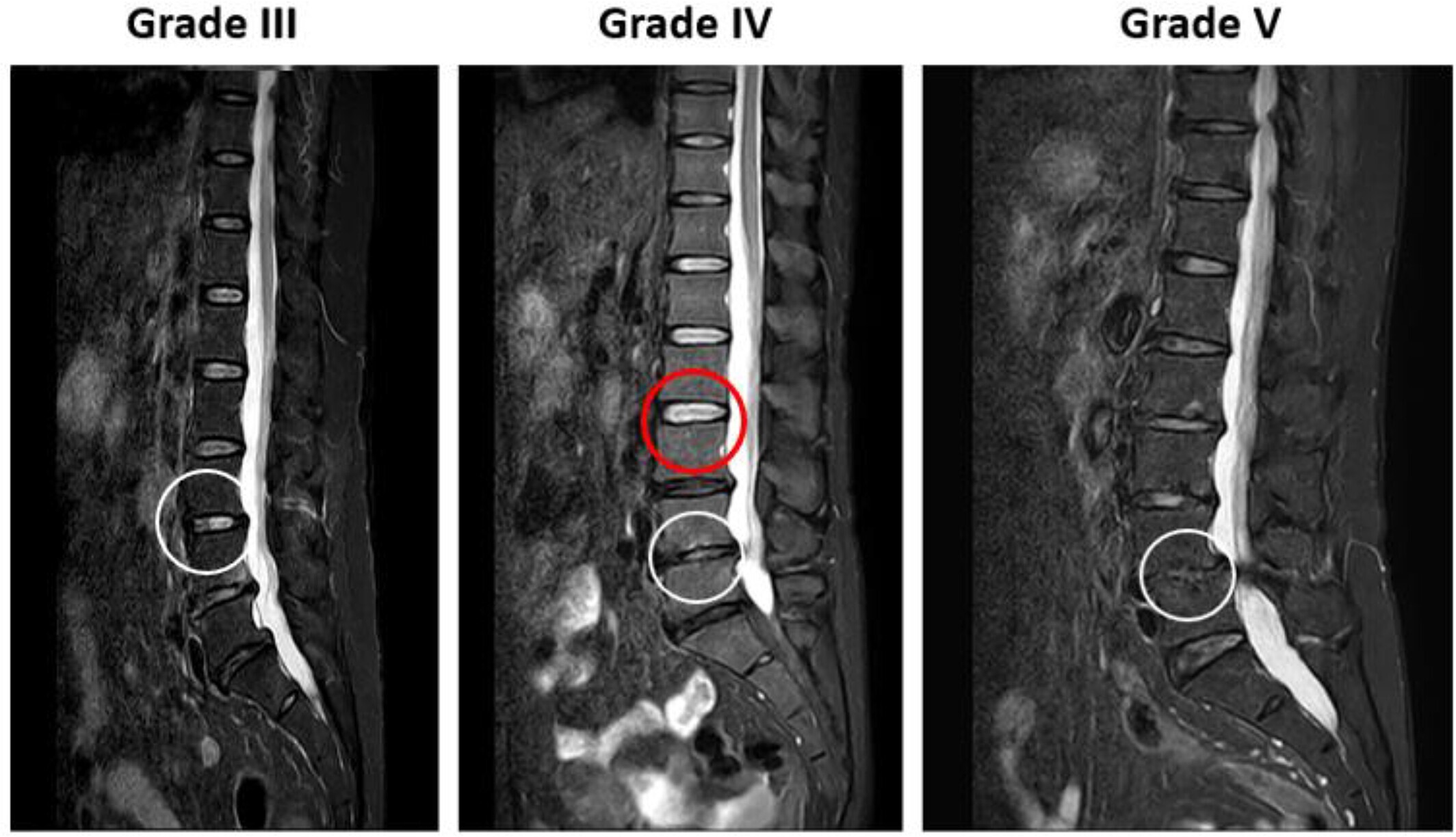
Representative magnetic resonance imaging scans of the degenerative disc based on the Pfirrmann grading system. The white circles indicate the location of degenerative discs, and the red circle is a healthy disc, which is referenced for comparison.
The collaboration between Dr. Kuo-Yuan Huang and Prof. Yeau-Ren Jeng originated from Dr. Huang’s observation that patients undergoing surgery have varying blood pressure levels. His initial idea was to develop a dynamic tourniquet system for knee replacement surgeries, allowing for blood pressure control tailored to each case, thereby reducing the occurrence of venous thrombosis. He then sought the expertise of Prof. Jeng, an authority in the field of tribology.
They concluded that Taiwan lacked precision instruments for assessing human body composition. As a result, Professor Jeng developed precision testing instruments to explore the different degrees of degeneration in intervertebral discs. This would involve examining the corresponding internal tissue components, as well as their viscoelasticity and biomechanical functions. Ultimately, this research would enable tailored treatment decisions based on individual test results.
Prof. Yeau-Ren Jeng stated that modern people often remain in a “text neck” posture by constantly looking down at their phones. Prolonged poor posture can lead to sore shoulders and neck pain. After age 30, blood circulation decreases, making individuals more susceptible to conditions like disc herniation and bone spurs—the fluid between joints functions like lubricating oil in a machine. With prolonged use, it gradually depletes. The vertebral joints’ cartilage can be likened to tires; the longer they are used, the more the surface thins, increasing the risk of accidents, which can be dangerous.
Like equipment, the human body also experiences wear and tear over time. To obtain an in-depth analysis of the internal condition of intervertebral discs, this innovation employed a system integrating nanoindentation with Raman spectrometry and incorporating artificial intelligence technology. This aids doctors in accurately assessing the degree of spinal degeneration.
Prof. Yeau-Ren Jeng explained that this technology can directly measure changes in the biochemical makeup of the intervertebral discs, allowing doctors to understand the state of their weight-bearing ability, whether there are issues like slippage or elastic degeneration, and to assess whether there are potentially dangerous situations like structural instability or nerve compression. This prior assessment allows further degeneration analysis with tiny tissue samples. This innovation is a crucial technique for tissue regeneration and cellular therapy. The team will combine this study with big data analysis to contribute to developing new drugs and intervertebral disc substitutes for spinal issues, improving patients’ quality of life and heralding a new era of precision medicine.
Provider:
NCKU News Center
Date:
2023-09-13
